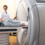
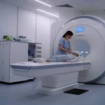
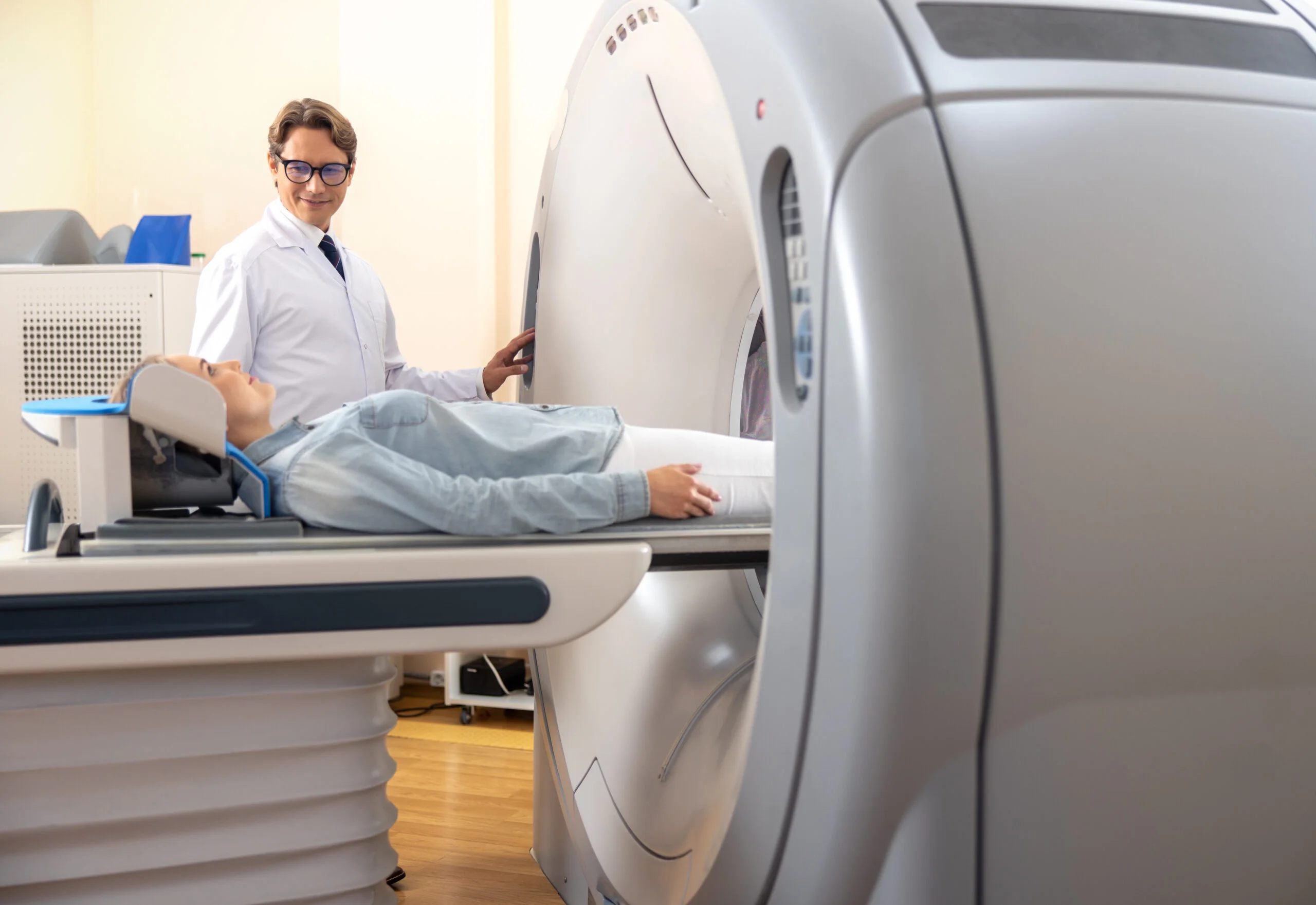
After your doctor has ordered an MRI, it isn’t difficult to find yourself pondering what on earth they’re staring at you for. Magnetic Resonance Imaging, or MRI, is famous for taking just fantastic photos of the interior of your body—but what’s the photo indicating? From muscles and joints to organs and brain, MRI scans offer such a splendid visual map that helps radiologists diagnose, monitor, and treat a plethora of health conditions. In this blog, we’re going to break down what an MRI image looks like, how it’s produced, and what different tissues and abnormalities may appear like on imaging center Glendale the scan.
What makes MRI images different from other scans?
MRI scans are special in that they are high-contrast and high-resolution, but particularly for soft tissues. In contrast to X-rays or CT, which are more suited to imaging center Glendale CA bone, MRI scans show anatomy of muscle groups, ligament, cartilage, nerves, and organs.
The MRI does this using no radiation. The MRI scans with strong magnets and radio waves to detect the signals from your body’s hydrogen atoms. Because your body is full of water and water contains hydrogen, different tissues emit differing signals based on structure and density. The signals are read off on gray-scale pictures, gradients of gray in order to delineate between differing tissues.
What is an MRI image composed of?
An MRI image is usually delivered in slices—flat, thin sections that uncover the body layer by layer. They can be seen from many angles: axial (side to side), coronal (back to front), or sagittal (front to back). Picture slicing a loaf of bread. A new angle is uncovered with every slice so that radiologists can observe issues in one location.
Everything is depicted in black and white or in shades of grays. Adipose tissues will be white, and more dense structures like tendons or fluid compartments will be darker. Tissue appearances will also depend on whether the scan was in which mode, i.e., T1-weighted or T2-weighted imaging. Different modes highlight various anatomical structures.
What does a given body region look like on an MRI scan?
White and gray are seen on a brain MRI. White is darker, and gray is lighter. Smaller structures slightly smaller, including cerebral cortex folds, ventricles (fluid spaces), and brainstem, are seen.
On a spine MRI, the spinal cord can be seen as it traverses the vertebrae. Herniation of the disc, spinal canal stenosis, and compression of nerves are all readily apparent as dark or light spots other than normal anatomy.
Shared MRIs, such as the knee or shoulder, highlight ligaments, cartilage, tendons, and muscles that encircle them. Those which are torn or ruptured will be evident from those that are intact. A meniscus tear, for instance, will be indicated by an abnormal curve or white line in the black area of the cartilage.
On a belly scan, the pancreas, kidneys, and liver are seen clearly. Tumors, cysts, or swelling are lighter to some degree, depending on the type of scan and if dye is injected to make them stand out so they can be seen.
How do MRI images reveal abnormalities?

The most powerful MRI imaging emphasizes differences in tissue density and structure. When something is amiss—a tumor, trauma, or infection, for example—the tissue signal is altered. Radiologists learn to recognize signal changes.
Tumors are seen as a region of brightening or darkening, depending on the type of scan. Edema or swelling is typically white on T2-weighted images. Infection, abscess, and hemorrhage are common findings. Contrast agents can be used to further enhance the visualization of these lesions.
Radiologists seek an asymmetry, abnormal radiolucency or radiopacity, and change in the texture of the tissue. These signs, combined, build a diagnosis.
Why are MRI scans typically black and white?
MRI scans are black, white, and gray because the information are recorded and processed in such a manner. Your body emits magnetic resonance signals, not color signals. Imaging software will sometimes overlay color maps to show differences between various types of tissue or activity, especially for functional MRI scans.
Although MRI isn’t painted with colors, contrast is incredibly, incredibly high. This indicates that doctors can recognize very small changes in tissue—more than on other imaging studies in certain situations. So you don’t have a Technicolor presentation, but what you have is gritty and diagnostic in tone.
Can patients read MRIs?
Most MRI scans resemble abstract art to the unaided eye. The silhouettes and shadows may be difficult to interpret in the absence of a radiology and anatomy experience. Nevertheless, with the guidance of a doctor, patients have a tendency to grasp the fundamentals. Your doctor can identify landmark structures, say what appears to be normal anatomy, and where there is something abnormal.
Radiologists create reports in great detail that take what is showing on the images and turn it into something tangible. Those are what your referring doctor presents to let you know what you should do next. Even though you may not have to interpret the scan completely, seeing what is showing on the picture can help frame the process within context.
How are MRI images stored and transmitted?
Once scanned for an MRI, images are captured in computer form as DICOM. Images can be stored in hospital systems, transferred securely to specialists, or posted on patient portals. The images are then read by radiologists, who then report back to the referring physician by summary and interpretation.
They even provide you with a copy in digital form in an imaging center Glendale. It will come in handy when you are searching for second opinions or if you have to make use of the services of multiple physicians. It will not show everything when you are viewing the images yourself, but it will keep you awake and alert to your medical care.
Do there exist different types of MRI pictures?

Yes. Clinical setting defines MRI Glendale imaging center Glendale ca. T1-weighted images are fat-sensitive and produce better anatomic data. T2-weighted images are fluid-sensitive and best for detecting edema or inflammation. Specialized ones do exist, such as diffusion-weighted imaging for detection of acute tumor or stroke and functional MRI, which detects localization of brain activation.
Each scan is different in contrast so that doctors are able to more clearly see what is occurring in the body. It is the difference that is making MRI so effective as a diagnostic tool.
Conclusion
An MRI scan may look like a black-and-white spiral to the naked eye, but to a radiologist, it is a very detailed picture of your inner world. From charting tumors and snapped ligaments to revealing neurological changes, MRI gives doctors the information they need—one piece of information at a time.